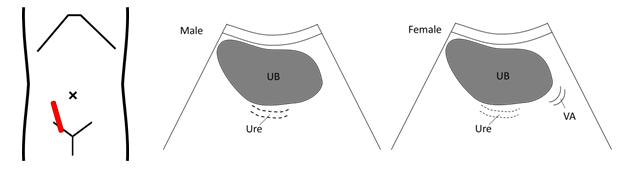
Pelvic Female Full Abdomen Ultrasound / Gynaecology Ultrasound Women S Ultrasound Melbourne : An ultrasound of the pelvis is typically used to look at the bladder, ovaries, uterus, cervix, and fallopian tubes (some of these are known as the female reproductive organs).
Pelvic Female Full Abdomen Ultrasound / Gynaecology Ultrasound Women S Ultrasound Melbourne : An ultrasound of the pelvis is typically used to look at the bladder, ovaries, uterus, cervix, and fallopian tubes (some of these are known as the female reproductive organs).. The abdominal, the pelvic/gynaecology and the urinary ultrasound scan.this ultrasound examination will examine all the abdominal organs visible on ultrasound scanning. Her physical exam revealed massive abdominal distention and spider angiomata suspicious for possible ascites. Complete pelvic ultrasound (upeltv) this is a complete pelvic ultrasound exam, including transabdominal and transvaginal. A hand held device called a transducer (also called a probe or wand) sends and receives these soundwaves. However subsequent ultrasounds may be ordered after that based upon your growth and condition.
Imaging tests can identify abnormalities and help doctors diagnose conditions. Having a full bladder can help with looking at organs, such as the womb (uterus), within your pelvis. Assessment of the pelvic floor can be accomplished with either us or mr imaging. A pelvic ultrasound is a test that uses sound waves to make a picture of the inside of the lower belly (pelvis). A pelvic ultrasound may be done with a full bladder.
Ultrasound Scans For Infertility Testing And Treatment from www.verywellfamily.com The abdominal, the pelvic/gynaecology and the urinary ultrasound scan.this ultrasound examination will examine all the abdominal organs visible on ultrasound scanning. Ultrasound abdomen is one of the tests that is commonly used in the assessment of patients with abdominal pain. An ultrasound of the pelvis is typically used to look at the bladder, ovaries, uterus, cervix, and fallopian tubes (some of these are known as the female reproductive organs). The sound waves create a picture on a video monitor. However subsequent ultrasounds may be ordered after that based upon your growth and condition. A pelvic ultrasound is a test that uses sound waves to make a picture of the inside of the lower belly (pelvis). Lower pelvic fullness and discomfort, bloating. You don't need to change your diet or avoid any particular foods.
However subsequent ultrasounds may be ordered after that based upon your growth and condition.
It's okay to drink caffeinated products prior to an ultrasound, so you can have coffee, soda, and other caffeinated drinks. I also had to urinate constantly (twice a night in my sleep). Transabdominal pelvic ultrasound can detect most larger abnormalities such as large fibroids, ovarian cysts, neoplasms, etc. A hand held device called a transducer (also called a probe or wand) sends and receives these soundwaves. Pelvic ultrasound can help your doctor identify problems in your lower abdominal and pelvic organs, such as your bladder. It allows your doctor to see your bladder, cervix, uterus, fallopian tubes, and ovaries. Other codes that can be used as needed are upelc, upell, utvag. Ultrasound imaging uses soundwaves to create pictures of the inside of the body. Transabdominally (through the abdomen) and transvaginally (through the vaginal canal). Lower pelvic fullness and discomfort, bloating. A pelvic ultrasound may be done with a full bladder. A pelvic ultrasound allows quick visualization of the female pelvic organs and structures including the uterus, cervix, vagina, fallopian tubes and ovaries. The sound waves create a picture on a video monitor.
It started as a dull, localized throbbing or tingling on my right side, but got worse with each day. A pelvic ultrasound is a test that uses sound waves to make a picture of the inside of the lower belly (pelvis). Rectal, or transrectal, ultrasounds can make images of a man's prostate gland and seminal vesicles. You should wait until after the test to urinate. Having a full bladder can help with looking at organs, such as the womb (uterus), within your pelvis.
Japanese Society Of Sonographers from www.jss.org A pelvic ultrasound scan can be performed in two ways: Transabdominally (through the abdomen) and transvaginally (through the vaginal canal). Assessment of the pelvic floor can be accomplished with either us or mr imaging. Rectal, or transrectal, ultrasounds can make images of a man's prostate gland and seminal vesicles. Find problems with the structure of your uterus or ovaries look for cancer in your ovaries, uterus, or bladder find an intrauterine device (iud) I also had to urinate constantly (twice a night in my sleep). Imaging tests can identify abnormalities and help doctors diagnose conditions. In women, doctors can use a pelvic ultrasound to:
Pelvic ultrasound can help your doctor identify problems in your lower abdominal and pelvic organs, such as your bladder.
Obstetric ultrasound second trimester begins starting from 14 weeks pregnant to about 28 weeks. A quick scan of the true and false pelves at the end of the study after the patient has voided is part of a routine examination. The sound waves create a picture on a video monitor. Transabdominal pelvic ultrasound can detect most larger abnormalities such as large fibroids, ovarian cysts, neoplasms, etc. Pelvic ultrasound getting a pelvic ultrasound ordered from your physician usually occurs because he/she is interested in seeing your uterus or ovaries. Having a full bladder can help with looking at organs, such as the womb (uterus), within your pelvis. And the use of ultrasound to diagnose pelvic pain in women. The types of pelvic ultrasound include: It started as a dull, localized throbbing or tingling on my right side, but got worse with each day. An abdominal ultrasound is a test that results in images of the upper abdominal viscera, or solid organs. A pelvic ultrasound can be performed at any stage of a woman's menstrual cycle. However, its views may be limited by abdominal structures such as bowel gas. In women, a pelvic ultrasound may be used for a number of reasons, including those listed below.
You should wait until after the test to urinate. Depending on the patient and the condition being assessed, either one or both of these methods can be used. Reasons for having an abdominal and pelvic ultrasound scan include: Go ahead and eat whatever you want. The sound waves create a picture on a video monitor.
Why You Shouldn T Empty Your Bladder When Preparing For An Ultrasound Independent Imaging from wp02-media.cdn.ihealthspot.com A pelvic ultrasound may be done with a full bladder. The two essential parts of a woman's female anatomy includes the uterus and the ovaries. You don't need to change your diet or avoid any particular foods. Other codes that can be used as needed are upelc, upell, utvag. Abdominal, or transabdominal ultrasounds can produce images of the bladder, uterus, cervix, ovaries and fallopian tubes. In the pelvic cavity, there are portions of the intestines, the organs of the reproductive, the urinary, and a large part of the musculoskeletal systems. An ultrasound of the pelvis is typically used to look at the bladder, ovaries, uterus, cervix, and fallopian tubes (some of these are known as the female reproductive organs). Pelvic ultrasound getting a pelvic ultrasound ordered from your physician usually occurs because he/she is interested in seeing your uterus or ovaries.
The uterus or womb is a pear shaped organ that can be seen in female patients of all ages.
The types of pelvic ultrasound include: An abdominal ultrasound is a test that results in images of the upper abdominal viscera, or solid organs. In general for pocus exams, it is usually good to start with the transabdominal ultrasound and then use the transvaginal approach if needed. Having a full bladder can help with looking at organs, such as the womb (uterus), within your pelvis. A pelvic ultrasound can be performed at any stage of a woman's menstrual cycle. A pelvic ultrasound is a test that uses sound waves to make a picture of the inside of the lower belly (pelvis). An abdominal ultrasound has no risks. Having a full bladder can help with looking at organs, such as the womb (uterus), within your pelvis. The two essential parts of a woman's female anatomy includes the uterus and the ovaries. However, the imaging test may be used to diagnose or rule out many other health conditions. An ultrasound of the pelvis is typically used to look at the bladder, ovaries, uterus, cervix, and fallopian tubes (some of these are known as the female reproductive organs). In women, doctors can use a pelvic ultrasound to: Ultrasound uses high frequency sound waves which pass through the skin and are reflected off the organs to create a picture on a screen with the help of a computer.
However subsequent ultrasounds may be ordered after that based upon your growth and condition pelvic ultrasound female. The sound waves create a picture on a video monitor.
/pregnancy-ultra-sound-97530915-588be9f05f9b5874eed3b0d2.jpg)


Komentar
Posting Komentar